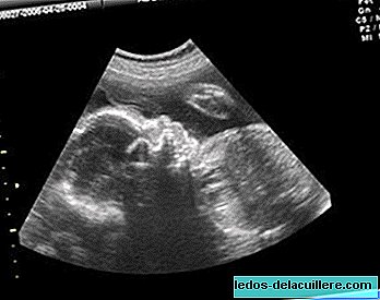
The ultrasound it's a technique that uses ultrasound waves which are reflected in the different tissues of the human body, and when they return to the transducer, they become signals that allow us to see images of these tissues on a screen.
As it is simple, harmless and cheap is the technique that is most used in prenatal tests for pregnancy monitoring. To perform it, the doctor slides a device called a transducer over the gut of the pregnant woman or introduces it through the vagina, depending on how advanced the pregnancy is.
Risks: safe for both the woman and the baby, as long as it is used properly and it is necessary to obtain medical information about the pregnancy. Casual use should be avoided during pregnancy. The woman feels pressure as the transducer moves but the procedure is not painful.
Several ultrasounds are performed during pregnancy, they must be a minimum of three, but in many cases they tend to be more.
- Between 8-14 week: transvaginal is performed. The main utility is to calculate the gestational age by measuring the skull-spinal length (CRL). It also rules out ectopic pregnancy, multiple pregnancy and malformations (assesses, among other things, the nuchal fold, ossification of the nasal bone, dopler of the ductus arteriosus) The baby's heartbeat can already be felt (from 7th week).
- Between 18-20 weeks: through trans-abdominal means. Especially for the assessment of possible malformations, and monitoring of gestational age through the measurement of biparietal diameter (BPD), femur length and abdominal diameter.
- Between 34-36 weeks: trans-abdominal, rule out a possible intrauterine baby growth delay
Performing this test during the second and third trimesters is the most effective way to estimate gestational age. This is achieved by measuring the diameter between the parietal bones of the skull and the length of the femur. We have to take into account that as the pregnancy progresses, it becomes more difficult to calculate it with ultrasound, and it may be in the last quarter with a variation of +/- 21 days.
Ultrasound in 3 dimensions or 4D (3 dimensions in movement) is the improvement in the image quality by projecting it in more dimensions, it gives a more real image of the fetus but is not more sensitive when discarding certain anomalies than the two-dimensional one.
Within the ultrasounds you can add the doppler technique, which is used to measure and evaluate the flow of blood that circulates through the circulatory system and the baby's heart and especially the umbilical arteries that provide oxygen to the fetus. It is indicated in high risk pregnancy. Measure the degree of fetal distress.
Apart from the medical point of view, it is the first contact parents have with the baby, they see him move, suck, sometimes even yawn, so it is also very important from the emotional point of view.
In Babies and more | Special pregnancy
In Babies and more | Ultrasound: all the news
Follow


